
Samsun Isthmocele Treatment
Samsun istmosel treatment involves the evaluation and guidance of scar defects in the post-caesarean uterine front wall according to symptoms and pregnancy plan.
Samsun istmosel treatmentIt includes the symptoms of scar defects in the uterine front wall after the caesarean section and their evaluation and guidance according to the pregnancy plan.
Istmosel, also called niche in medical language, is the pocket or tunneling that occurs on the front wall of the uterus as a result of the C-section not healing fully. A previous C-section is considered a late-term side effect of childbirth and does not follow the same severity in each patient. The size, location and thickness of muscle tissue in the uterine wall are among the factors that directly affect the type and severity of complaints that may occur.
Some ismosel cases give no symptoms and are randomly identified, in which case treatment may not be required and regular monitoring may be seen as sufficient. In patients with obvious complaints, it is important to evaluate the condition in detail by a physician. Prof. Dr. Aşkı Ellibeş Kaya Based on the results of the examination and imaging performed by the patient, monitoring or surgical options are handled together with the patient's overall condition.
What is an Istmosel (Caesian Skar Defective)?

Istmosel is a gap that occurs during the cesarean delivery as a result of the fact that the incision to the uterine front wall does not close completely in the healing process. This gap can be in the form of a small notch, as well as a deeper pocket or tunnel view. The size and depth of the defect vary from person to person, so not every istmosel forms the same clinical table, and the assessment is tailored to the individual. Although different rates are reported in the literature about the frequency of this defect, the detection rate is increasing thanks to current imaging methods.
As the number of caesareans increases, it is also considered to increase the likelihood of istmoceles being seen; Only after a single caesarean section can the defect develop. The position of the uterus, the level at which the precision is made, and the individual differences in the healing process are counted among the factors that play a role in shaping the defect together.
The clinical significance of istmosel is not limited to the complaints it causes only; When a planned pregnancy is involved in the future, the location and depth of the defect become a separate consideration. Therefore, it is recommended that patients with istmocele be registered and followed up when necessary, even if there is no apparent convergence.
What Are the Symptoms of Istmosel?

Istmosel does not always give symptoms; Small defects are often noticed by chance, for example during ultrasound for another reason, and may not cause any complaints. In obvious defects, symptoms such as postmenstrual and dark spotting, pain during intercourse with an increase in menstrual pain may be prominent. The severity of the findings may vary depending on the size of the defect and its position in the uterine wall.
Postmenstrual prolonged, dark spotting
Significant increase in menstrual pain (dismenorrhea)
Pain during intercourse (disparoni)
Secondary infertility in some cases
Blood accumulated in the defect and the outbreak is thought to make it difficult to pass into the cervix, making it difficult to conceive in some patients. However, its istmocele presence does not necessarily mean a diagnosis of infertility in itself; The evaluation of infertility, along with other factors, should be dealt with in a holistic approach.
The severity of the symptoms may change over time; In some patients, the findings, which are mild in the first years, may become apparent in the following period. It is recommended that patients who notice a permanent change in their menstrual pattern, experience recurrent pain during intercourse, or have difficulty conception, plan a gynaecology examination by specifying the C-section story.
In cases where intimidations significantly affect the quality of daily life, it is recommended to schedule a gynaecology examination without wasting time. Early evaluation makes it easier to both clarify the diagnosis and determine the appropriate monitoring or treatment option.
Why Istmosel Forms?
The most important reason for istmocele is that a sufficiently strong tissue repair cannot be achieved during the recovery process of the caesarean incision. Factors such as the technique of closure of the precision, the layout of the seam layers, and the capacity for tissue healing can play a role in the formation of the defect. Having had more than one caesarean section is considered to be among the factors that increase risk.
The stage of progression of the birthing act, the level at which the incision is opened in the uterus, and the conditions that make it difficult to recover after surgery are also among the factors associated with defect formation.The combination of these factors can increase the likelihood of istmocele development.
Chronic conditions affecting maternal age, general health status, and tissue healing capacity are also evaluated among the factors that can indirectly affect istmocele formation. In addition, the short duration between successive pregnancies can lead to a new surgical procedure without adequate recovery of the uterine wall.
The sewing technique used during the caesarean and the number of layers where the precision is closed are also among the technical elements associated with the risk of istmosel. Therefore, the surgical technique is considered an important variable that affects whether or not defecation occurs.
How to Get an Istmosel Diagnosis?

Istmosel diagnosis is supported by imaging methods, taking into account the patient's complaints and the history of the C-section. During the examination, the physician tries to assess both the presence of the defect and its possible size and depth; This assessment forms the basis for the subsequent treatment decision. The diagnostic process is usually completed from the foot and does not require additional preparation.
Transvaginal Ultrasonography
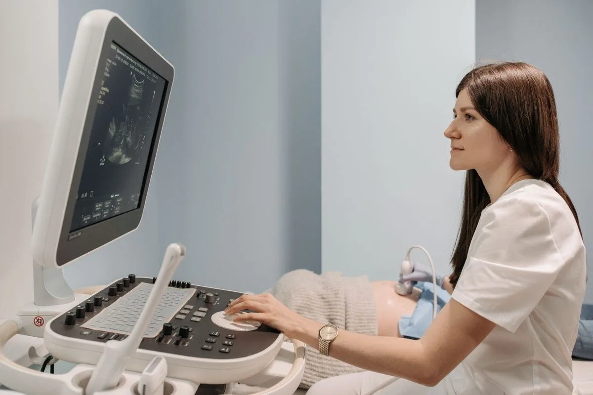
The first step in diagnosis is transvaginal ultrasonography. This method examines the incision line on the uterine front wall, if any, the size, depth of the defect, and the ratio to the uterine wall. Most istmosels are noticed during this review, and the initial evaluation is done in this way.
Salin Infusion Sonography
Infusion sonography of saline may be preferred when the boundaries of the defect need to be displayed more clearly. In this process, ultrasound image is clarified by giving a small amount of sterile fluid into the uterus and the shape of the defect, its depth and its relationship with surrounding tissues are evaluated in more detail.
Imaging results reveal not only the presence of the defect, but also the thickness of tissue that remains in the uterine wall. This information is important data to guide the physician as to whether monitoring or surgical repair will be recommended.
Istmosel Treatment Options
The treatment approach is shaped by the severity of the symptom caused by the istmocele and the patient's pregnancy plan. While monitoring may be sufficient in mild cases without the need for any attempt, surgical repair options may be raised in cases with significant complaints or associated infertility.
Before the treatment decision is made, the patient's previous caesarean section, the duration of their current complaint, and the future pregnancy plan are questioned in detail. This information plays a decisive role in shaping the preference between monitoring and surgical repair.
Monitoring and Hormonal Treatment
Regular monitoring may be seen as sufficient in the istmosel, which does not give symptoms or is mildly observed; a control examination is recommended at certain intervals in this approach. In cases where bleeding complaint is at the forefront, hormonal treatment options for controlling bleeding may be evaluated by the physician.
Hysteroscopic Repair
In hysteroscopic repair, the edges of the defect are corrected by entering the uterus, and the incision line is intended to gain a smoother appearance. This method is usually an option that is assessed in more superficial defects, in cases where uterine wall thickness is sufficient.
Laparoscopic Repair
Laparoscopic repair can be evaluated in deeper defects or when the uterine wall is noticeably thinned. In this method, the defected tissue is removed and the uterine wall is re-repaired layer by layer and the wall thickness is aimed to increase.
Which method to choose is not a single criterion; The properties of the defect are based on the evaluation of multiple factors, such as the patient's complaints and the pregnancy plan. Therefore, a detailed examination and imaging process is recommended prior to the treatment decision; Sharing the patient's questions and concerns with his or her physician contributes to the decision process.
Differences Between Treatment Methods
The following list compares the three approaches that are frequently used in istmosel management to their general outline, which method is appropriate, examination findings, depth of defecation and the patient's pregnancy plan are evaluated together and determined by the physician.
No surgical intervention is applied in the follow-up approach; The patient is called to a control examination at certain intervals. In surgical repair options, hysteroscopic and laparoscopic methods aim to correct the defect with different access techniques, and the choice is made according to the characteristics of the defect.
Watch: It is maintained at certain intervals with a control examination and, if necessary, hormonal support; it is usually assessed in defects that do not show symptoms or are mildly observed.
Hysteroscopic Repair: The edges of the defect are corrected by an attempt from within the uterus; It is an option assessed in superficial defects and in cases with significant bleeding complaints.
Laparoscopic Repair: A defected tissue is repaired by an attempt from inside the abdomen; It is evaluated in deep defects, cases where the uterine wall has been thinned and cases with a pregnancy plan.
There can be no precise ranking of superiority among the approaches listed; Each is an option for a different clinical table. In the decision phase, the patient's complaints, imaging findings and future pregnancy plan are considered together.
Istmosel and Pregnancy Planning
In patients planning pregnancy in the future, it is generally recommended that istmocele repair be performed before pregnancy. Istmoselin is known to be rarely associated with serious complications in future pregnancies. Therefore, pre-pregnancy assessment is considered an important part of the treatment decision.
Patients with a pregnancy plan, a Samsun Gynaecologist It is recommended that it be evaluated both in terms of the condition of istmocele and overall reproductive health. This assessment can contribute to safer planning of the subsequent pregnancy process and to prior realization of possible risks.
If a new pregnancy is planned after repair, it is usually recommended to wait for a period set by the physician to complete the recovery process. This time can vary depending on the method applied and the patient's recovery rate; The exact time is assessed during the inspections.
It should be remembered that the repair decision can also be evaluated independently of the pregnancy plan; In some patients, surgical repair may be preferred only for the purpose of eliminating complaints. In this case, the decision is made taking into account the patient's priorities and examination findings together.
Is It Possible to Protect from Istmosel?
While there is no method that completely eliminates the risk of istmosel, some factors can be mentioned that may reduce the risk. These factors include reducing the number of unnecessary caesareans and the proper surgical technique of the caesarean incision, layer by layer and carefully closed. This approach may contribute to the more robust recovery of the incision line.
Regular follow-up of the postoperative recovery process and failure of control examinations when necessary can also help to detect a possible defect early on. This approach facilitates earlier detection of complaints that may arise in the future.
The decision of the caesarean is considered an important approach for both maternal and uterine wall health, only in the case of medical necessity. Detailed consultation with the physician on the form of childbirth may contribute to the prevention of late-term problems such as istmoceles in subsequent pregnancies.
Making the first post-caesarean check-up on time allows for close monitoring of the healing process. This follow-up is considered a valuable habit in terms of early detection of complaints that may arise in the future.
How Does the Istmosel Evaluation Process Work?
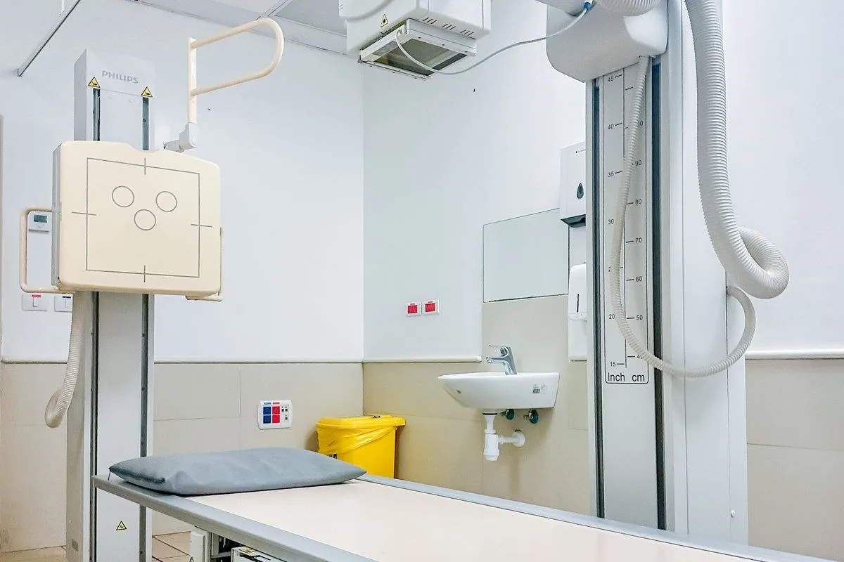
The evaluation process usually begins with the rest of the patient's complaints, gynecological examination and imaging methods. A road map is created between monitoring or surgical repair options, with the size, location of the defect and the patient's pregnancy plan being discussed together. The process may not result in a single examination and control appointments may be scheduled if necessary.
The main elements that the physician takes into account during the evaluation include:
The size and depth of the defect
The severity of the patient's complaints
Number of caesareans passed
Whether there is a pregnancy plan
Each of these elements directly affects the preference between monitoring and surgical repair. A detailed briefing to the patient explaining the current situation and possible options as a result of the evaluation is an important step that facilitates its participation in the treatment process.
Caesarean Timing in the Next Pregnancy
When a subsequent pregnancy occurs in patients with an istmosel history, it requires a separate assessment of when and by which method the birth will be planned. The integrity of tissue in the previous incision line is a re-emergence factor with the strain of the uterine wall as the pregnancy progresses. Therefore, the frequency of follow-up and imaging plan in pregnant women with istmosel history may vary compared to pregnant women with no history.
The main factors observed by the physician in caesarean timing include the thickness of the uterus wall, the nature of previous repair, and the findings identified during pregnancy. Rather than waiting for spontaneous labor to begin, the preference of planned caesarean during a designated pregnancy week may be seen as more appropriate in some cases; This approach aims to make the incision line less exposed to the stress process. However, this decision is not taken in the same way in each patient, and the course of pregnancy and examination findings are evaluated together.
Ultrasonographic follow-up during pregnancy allows for monitoring of tissue thickness in the incision line and early detection of a possible thinning. This information guides the physician in what week and by which method the birth will be performed. Patients with an istmosel history are advised to continue their pregnancy follow-up with a physician who knows this story and to create birth planning together in accordance with these assessments.
Conclusion
Samsun istmosel treatment The approach determined in the process is shaped according to the patient's complaints, the characteristics of the defect, and the pregnancy plan. Different options, from monitoring to surgical repair, are discussed together after a thorough examination and imaging evaluation.
Patients who notice symptoms related to ismoselle or are curious about their past C-section history are advised to clarify their condition by consulting with a specialist. Early evaluation can contribute to healthier management of both current complaints and future pregnancy plans.

Original Turkish content prepared and reviewed by
Prof. Dr. Aşkı Ellibeş Kaya
Specialist in Obstetrics and Gynecology
Last updated: 23 August 2026
The content on this page is intended for informational purposes; the diagnosis and treatment decision is made only with your physician after the examination.
The information on this page is for general informational purposes only and does not replace medical advice, diagnosis or treatment. Each patient's condition is different; the method and process to be applied is determined only by the physician after examination and evaluation.
Frequently asked questions
Oh, no. In the istmosel, which does not show symptoms or is slightly visible, monitoring is often sufficient; Surgical repair is assessed in cases with significant complaints or associated infertility. According to the findings of the decision, examination and imaging, the patient is given in private by the physician.
Related Services
Other topics under the topic of gynecology.



Start with an appointment
Let us determine the day and approach that best suits you. Your consultations are conducted on a privacy basis.