

Samsun Uterine Polyp Treatment
Polyps are benign formations that develop from the inner layer of the uterus. They can have an effect on bleeding and fertility.
Samsun uterine polypy treatment, covers the diagnosis and treatment process of tissue growths resulting from the inner layer of the uterus. Early evaluation is recommended in the bleeding complaint.
uterine polybi is a growth of tissue that is often considered benign, resulting from the endometrium; The number may be one or more, and the size may vary significantly from person to person.
As it can be noticed by symptoms such as irregular spotting, intermenstrual bleeding or postmenopausal bleeding, it can sometimes also be detected coincidentally during a routine examination without causing any complaints; in some cases it is raised during a infertility assessment.
Ultrasound and hysteroscopy are used together in the diagnostic process; The precise diagnosis and treatment are usually planned together in the same session, aiming for the patient to receive results without the need for a second procedure. This evaluation process in Samsun Prof. Dr. Aşkı Ellibeş Kaya It is conducted by examination, imaging and, when necessary, hysteroscopy steps.
What is uterine Polibi?

The uterus polybi is a tissue growth resulting from the endometrium layer covering the inner surface of the uterus. It is often considered benign and develops in the form of a soft tissue mass that extends into the uterine cavity, holding on to the stem or broad base.
The number of polyps can be one or more; Its size can also vary from a few millimeters to several centimeters. This difference can directly affect the severity of the complaints and the treatment approach that the physician will follow.
Some polyps cover a small portion of the uterine cavity, while others can spread over a wider area or settle near the cervix. Magnitude and settlement are considered to be important factors affecting both the emergence of complaints and the approach the physician will take.
The uterus polybi is structurally different from other benign formations such as myoma caused by the uterine muscles; The myoma develops from muscle tissue, and the polyp develops from endometrium surface tissue. This distinction is carefully performed by the physician in ultrasound and hysteroscopy assessment and directly affects the treatment approach.
Polyps of similar structure can be seen in the cervix, except for the cervix; But the subject of this article covers polyps caused by the endometrium, within the uterine cavity. Both settlements are evaluated separately in the examination and treated separately if necessary.
Why is uterine Polibi Formed?
No single exact cause has been identified in the formation of uterine polybi; The response of endometrium tissue to hormonal stimuli is thought to play a role in the process. The stimulating effect of estrogen level on tissue is cited as one of the topics that is often explored in this regard.
Older age, non-birth, and some chronic health conditions are among the factors associated with polyp development. However, the presence of these factors does not always mean that polyps will form; Each patient's history and examination findings are evaluated together to determine a specific approach to the person.
It is known that in women who use some hormone-containing treatments for a long time, endometrium tissue can respond more frequently to such stimuli. Therefore, it is important that women with such a history of treatment do not interfere with their regular gynecologic control, in terms of early detection of a possible polybin.
Being overweight is also counted as one of the factors that can affect hormone balance, and in this respect it can be associated with polyp development. However, the presence of such factors does not provide a definitive prediction; each patient's condition is evaluated separately, along with findings of examination and imaging.
What Are the Symptoms of uterine Polybi?
The uterus polybi can be noticed by chance during a routine gynecologic examination or ultrasound for some other reason, without causing any apparent complaints in many women. In some cases, various bleeding irregularities are prominent and cause the patient to undergo an examination.
Irregular staining seen during menstruation
Bleeding that occurs between menstrual periods
Bleeding seen during the postmenopausal period
Period bleeding lasts longer than expected or increases in quantity
Detection during infertility assessment in some cases
Spotting that can occur after sexual intercourse
Coincidental detection in routine examination without any symptoms
The severity of the symptoms is closely related to the size of the polybin and its placement within the uterus; polyps that are located away from the center of the small and uterine cavity may remain undetected for a longer period of time, whereas polyps that are large or close to the cervix tend to cause earlier complaints.
When any of these symptoms are noticed, it is recommended to have a gynecological evaluation done without wasting time. Especially postmenopausal bleeding is considered a finding that must be evaluated regardless of the amount and the cause.
uterine Polybi Diagnostic Methods

The first step in the diagnosis process of uterine polybi is usually ultrasonography; When suspicious finding is detected, evaluation is supported by additional methods that clarify the image. Definite diagnosis and treatment are performed together in the same session, often accompanied by hysteroscopy.
Ultrasound and Salin Infusion Sonography
In standard ultrasound, thickening or mass image in the uterine inner layer may suggest polyp suspicion, but not always a definite distinction. With saline infusion sonography, the image is clarified by giving a small amount of fluid to the uterine cavity; This method contributes to a clearer view of polybin boundaries, number and settlement.
Definite Diagnosis with Hysteroscopy
Hysteroscopy is the method that allows for direct imaging of the inside of the uterus with the help of a thin camera, and is considered the gold standard in the diagnostic process. During this procedure, the polyp is directly observed, taking it in the same session if necessary, providing both diagnosis and treatment together.
None of the methods used in the diagnostic process may yield conclusive results alone; Therefore, the physician evaluates ultrasound findings along with the patient's complaints and examination results. Direct imaging with hysteroscopy when a suspicious image is detected is recommended for clarity of diagnosis.
Separator Evaluation
Since there are other reasons that can lead to abnormal bleeding, the physician also considers the possibilities outside the polyp during evaluation; Myoma, endometrium thickening and hormonal irregularities are part of this distinctive assessment. This approach contributes to the correct diagnosis and proper treatment planning.
Preparation Before Hysteroscopic Polypectomy
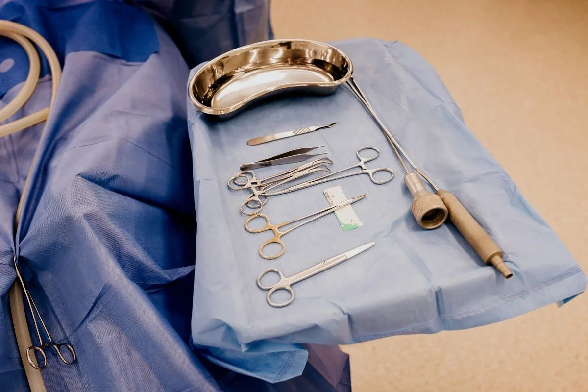
Prior to the procedure, the physician questions the patient's menstrual cycle, the medications he or she uses, and the history of bleeding disorders if any; This information directly affects process timing and anesthetic preference. The required blood tests and imaging may be requested as part of the process planning.
The procedure is usually planned by aligning it with the period at which the menstrual bleeding ends, as the inner layer of the uterus can be assessed more clearly during this period. It is important to inform the physician in advance about the use of drugs that may increase bleeding, in order to plan the procedure safely.
Comparison of Impression and Hysteroscopic Polypectomy in uterine Polypin
Not every uterine polybi is necessarily a finding that needs to be removed; Polyps that are small in size and do not cause complaints can be followed by monitoring at certain intervals in some cases. However, this decision is always made in person with physician evaluation, taking into account the age and complaints of the patient.
When monitoring is preferred, the follow-up is usually maintained by ultrasound checks performed at certain intervals; These controls assess whether there is a change in the size of the polybin and whether a new complaint has developed. If a complaint is filed during the follow-up, the approach will be reconsidered.
Status: Watch ; Hysteroscopic Polypectomy
Size: Small, millimeter size; Large or incremental size
Complaint: No symptoms; Complaint of bleeding or staining
Period: Reproductive age, low risk profile; Postmenopausal period
Approach: Ultrasound follow-up at certain intervals; Direct extraction and pathological examination
It is usually recommended to remove it for polyps detected in the postmenopausal period and polyps accompanying the bleeding complaint; Because there is rarely a malignant change within the polyp. This risk is considered to be one of the most important factors affecting the monitoring decision and shaping the physician's treatment recommendation.
Has been detected in the postmenopausal period
Accompanying the complaint of bleeding or staining
Size increases over time
Consideration that polybin may play a role in infertility assessment
Detection of suspicious appearance on ultrasound
How is a hysteroscopic polypectomy performed?

Hysteroscopic polypectomy is a procedure by placing a thin camera inside the uterus, allowing the polybin to be seen directly and removed from its source. This method is preferred over other approaches, as it allows the polybin to be separated from the intact tissue and removed fully.
The procedure is usually completed in a short time, under outpatient treatment; The patient can return to his or her daily life in a short time by being discharged on the same day. The application can be performed with general or local anesthesia, depending on the situation; The preference for anesthesia is determined by the physician according to the patient's condition.
The polyp is removed by direct monitoring thanks to the camera image; This allows the process to yield more reliable results compared to methods made to the blunt. Since the entire cervix can be assessed in the same session, if there is more than one polyp, they can all be taken in a single procedure.
Thanks to the fine tools used during the procedure, there is usually no need to overextend the cervix; This contributes to the more comfortable process of recovery. The received tissue is separated to be sent completely to the pathology laboratory at the end of the procedure.
Post-processing Process and Pathology Result
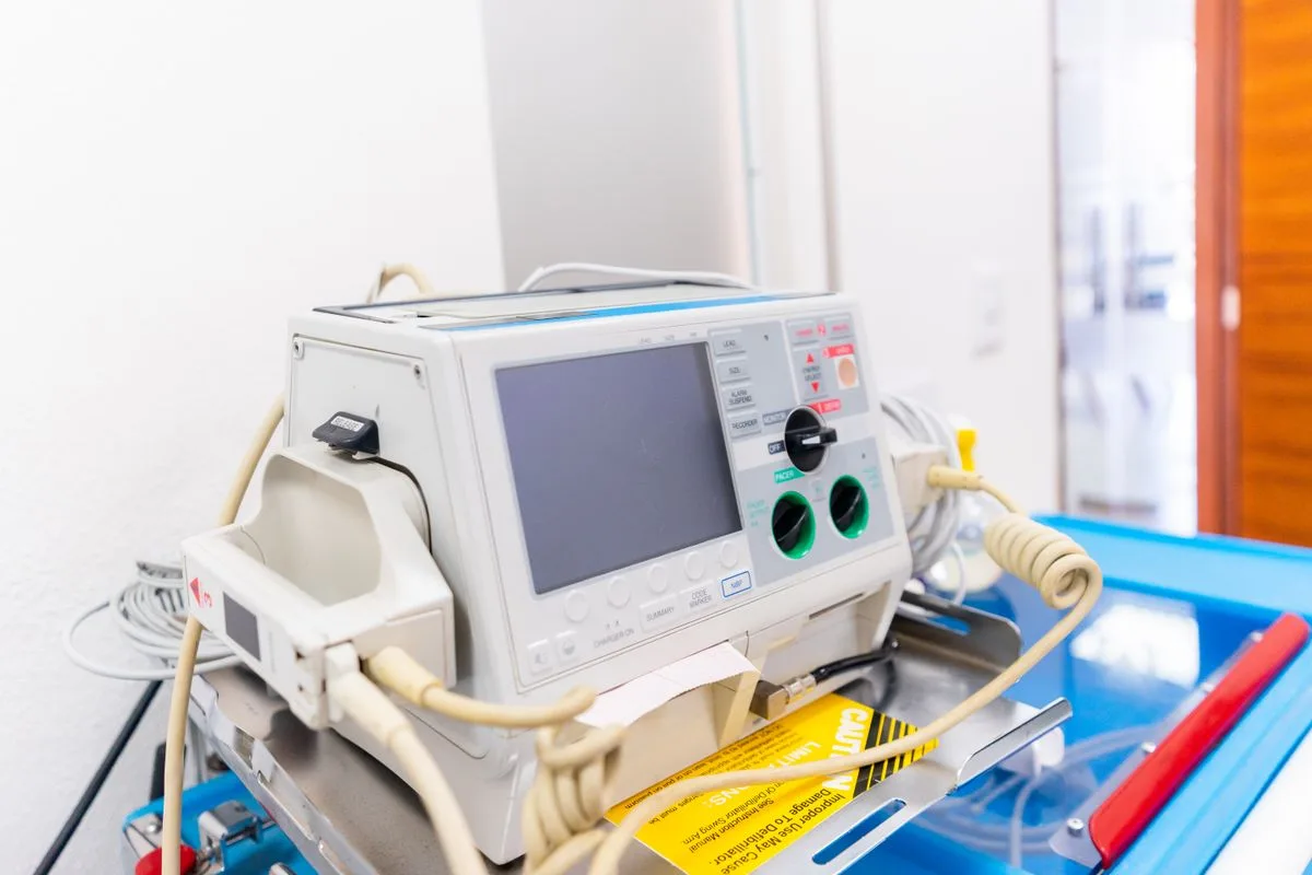
The tissue sample taken after the procedure is sent to the pathology laboratory; This review is important to clarify the polybin structure and plan an additional step if deemed necessary. The result is usually ready between a few days and a few weeks.
When the result comes out, the physician evaluates the findings with the patient and, if necessary, shapes the follow-up plan accordingly. In most cases, the result indicates benign changes and no additional treatment is needed.
After the procedure, mild spotting lasting for several days or feeling of mild cramping under the abdomen is considered normal. These symptoms usually go away in a short time by spontaneously decreasing; It is recommended to consult a physician in case of severe pain, prolonged bleeding or high fever.
It is often recommended that heavy exercise and strenuous physical activity be delayed for a few days following the procedure; This period may vary from person to person and the type of anesthesia administered. The physician informs the patient about specific recommendations after the procedure during discharge.
In matters such as sexual intercourse and tampon use, the physician may also recommend a period of respite until the complete end of staining. These recommendations are intended to ensure smooth completion of the healing process and reduce the risk of infection.
Polip Repetition Risk and Follow-up

After removal of the uterus polybi, it may re-develop in some cases; This can be especially common in people where hormonal factors persist or where endometrium tissue tends to do so. The likelihood of recurrence can be noticed in the early stage by regular follow-up.
Additional evaluation may be required in recurrent cases; The physician determines the frequency of follow-up and, if necessary, a new process plan, taking into account the previous pathology outcome and the course of complaints. Regular gynaecological controls are an important part of the process.
Since the frequency of recurrence may vary from person to person, it is important to follow the control intervals recommended by the physician after the procedure. When a new bleeding complaint arises, the assessment of this condition without delay makes it easier to spot a possible recurrence early.
Some patients may develop a new polyp in a different region later in the period, even if more than one polyp has been removed in the same session. Therefore, it is recommended that women with a history of polyp in the past maintain their routine gynecologic control at regular intervals.
uterine Polipi Evaluation in Samsun
The evaluation process for women with suspected uterus polybi or bleeding complaints begins with a thorough examination and the necessary imaging methods. One Samsun Gynaecologist The source of the complaint can be clarified by examination and, if necessary, the further examination process can be planned.
Following the evaluation, it is decided whether follow-up or hysteroscopic polypectomy is appropriate, the age of the patient, complaints, polybin imaging findings are taken into consideration together. The process is shared with the patient at each stage, and questions are answered.
The story and examination findings taken at the first application are completed with additional imaging when necessary, providing the basis for planning the next step. This approach allows both follow-up and hysteroscopic polypectomy decision to be shaped according to the patient's specific condition.
Note when the patient's complaints started before the appointment, their menstrual pattern and previous gynecologic evaluations if any can help the examination pass more efficiently. This information contributes to the physician's correct assessment.
Conclusion
Samsun uterine polypy treatmentA process that involves accurate recognition of tissue growths from the inner layer of the uterus and safe removal when necessary.The decision of a follow-up or hysteroscopic polypectomy is determined by the physician according to the patient's clinic and examination findings.
It is especially important that symptoms such as postmenopausal bleeding are evaluated without neglect or wasting time. Regular gynecological controls contribute to the early detection and proper management of a possible polybin.

Original Turkish content prepared and reviewed by
Prof. Dr. Aşkı Ellibeş Kaya
Specialist in Obstetrics and Gynecology
Last updated: 28 August 2026
The content on this page is intended for informational purposes; the diagnosis and treatment decision is made only with your physician after the examination.
References
The following sources relate to the informational issues outlined by them. The personal diagnosis and treatment plan requires physician evaluation. Source texts are in English.
NHS
Hysteroscopy (opens in new tab)Related subject: Evaluation and removal of polyps by hysteroscopy.
The information on this page is for general informational purposes only and does not replace medical advice, diagnosis or treatment. Each patient's condition is different; the method and process to be applied is determined only by the physician after examination and evaluation.
Frequently asked questions
uterine polybi is mostly a benign tissue growth; However, in rare cases, malignant change can be seen within the polyp. Therefore, the tissue removed is always sent to pathological examination and the result is shared by the physician with the patient.
Related Services
Other topics under the topic of gynecology.



Start with an appointment
Let us determine the day and approach that best suits you. Your consultations are conducted on a privacy basis.