
Samsun Hysteroscopy
Samsun hysteroscopy involves direct imaging of the inside of the uterus with a thin camera. It is used in abnormal bleeding, infertility research and intrauterine structural problems.
Samsun hysteroscopy with an intrauterine thin camera to be closely examined; No incisions are required, diagnosis and treatment can often be completed in the same session.
Hysteroscopy is an imaging method based on the delivery of an illuminated thin camera called a hysteroscope through the cervix. Abnormal bleeding is considered a reliable diagnostic and treatment tool that is frequently used in the evaluation of infertility research and intrauterine structural problems. Its ability to be applied without incision is also an important feature that facilitates the healing process.
Prof. Dr. Aşkı Ellibeş Kaya The evaluation by the nature of the complaint, examination findings and ultrasound results are taken into consideration together to determine whether hysteroscopy is necessary. This article discusses topics that are curious from the application of the method to the recovery process, from possible risks to frequently asked questions.
What is Hysteroscopy?
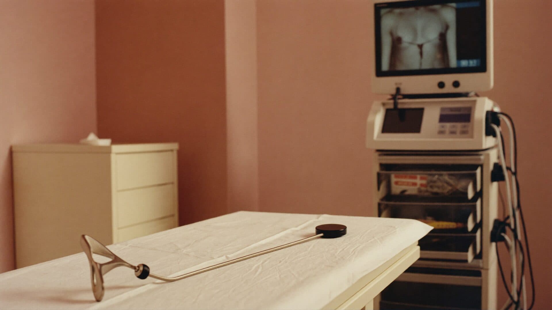
Hysteroscopy is a method of direct imaging of the inside of the uterus with a thin, illuminated camera called a hysteroscope. The camera is delivered through the mouth of the vagina and uterus into the uterus; This way, there is no need for any incisions in the abdomen. The image is projected on a monitor and evaluated in detail in the womb.
The method can be used for both diagnosis and treatment. In diagnostic hysteroscopy, the goal is to display the structure of the uterus and possible findings; In operative hysteroscopy, a problem identified can be treated in the same session. This feature makes hysteroscopy a practical method in gynecological evaluation.
Hysteroscopy can be performed in office conditions in some cases, as it can be applied in the operating room. Which environment is appropriate is determined by the scope of the planned procedure and the patient's overall condition. This flexibility allows the method to be adapted to different needs.
Ultrasound and other imaging methods provide indirect information about the intrauterine, while hysteroscopy provides direct observation. This direct imaging gives a significant advantage in clarifying the nature of a suspicious finding and, if necessary, in sampling tissue in the same session.
Detailed evaluation of an early-received finding with hysteroscopy helps to make subsequent treatment decisions more accurate, so it is worthwhile for women with complaints to apply for the examination without delaying evaluation, both in terms of diagnosis and possible treatment process.
How is hysteroscopy performed?
Pre-Process Preparation
A detailed gynecological examination is performed prior to the procedure and, if necessary, an ultrasound will complete the pre-assessment of the uterus. The physician receives information about the medications used, past surgeries and general health status. In some cases simple blood tests may be requested before the procedure.
Days near the end of the menstrual period are usually planned according to this period, as it allows for clearer viewing of the inside of the uterus. The physician determines the most appropriate date according to the patient's menstrual pattern and sets the date according to the patient's daily schedule if necessary.
Process Stages
During the procedure, the intrauterine is expanded with a sterile liquid or gas to clarify the image. The hysteroscope is passed through the cervix and moved into the uterus, and the camera image is monitored simultaneously. At the diagnostic stage, findings are evaluated; If operative processing is required, it is applied with the appropriate tools in the same session.
During the process, the image can be examined by magnifying, so that even small polyps or consonants can be evaluated in detail. The fine tools used in the operative stage are delivered to the uterus via the same hysteroscope, allowing the process to be completed in a single session.
Difference Between Diagnostic and Operative Hysteroscopy
Hysteroscopy is basically applied for two purposes: diagnostic and operative. In diagnostic hysteroscopy, the goal is to investigate the cause of abnormal bleeding, displaying anomalies of polyp, myoma, adhesion or uterine structure. In operative hysteroscopy, the detected finding is treated with the appropriate instruments in the same session.
Purpose: In diagnostic hysteroscopy, the cause of the complaint is investigated by displaying intrauterine; In operative hysteroscopy, procedures such as polyp removal, myoma removal or opening of adhesion are performed in the same session.
Media: Diagnostic hysteroscopy is usually performed in office conditions, while operative hysteroscopy is often preferred in the operating room environment.
Anesthesia: Local anesthesia is used in diagnostic hysteroscopy or no anesthesia is administered at all; In operative hysteroscopy, general or regional anesthesia is preferred.
Duration: Diagnostic hysteroscopy usually lasts for a few minutes, while the duration of operative hysteroscopy may be longer, depending on the extent of the finding detected.
Deportation: After diagnostic hysteroscopy, discharge occurs on the same day; In operative hysteroscopy, patients are often discharged on the same day.
The main difference between the two approaches is the scope of the process and the level of anesthesia it requires. Examination findings and previous examination results are determined by evaluating which method to apply; A finding that is often detected at the diagnostic stage can turn into operative processing.
In some patients, diagnostic hysteroscopy is planned first and after the findings are evaluated, it is decided whether the operative procedure is necessary. This step-by-step approach can also make it possible for the patient to receive the treatment he or she needs in a single session while preventing unnecessary procedures.
In What Situations Is Hysteroscopy Recommended?
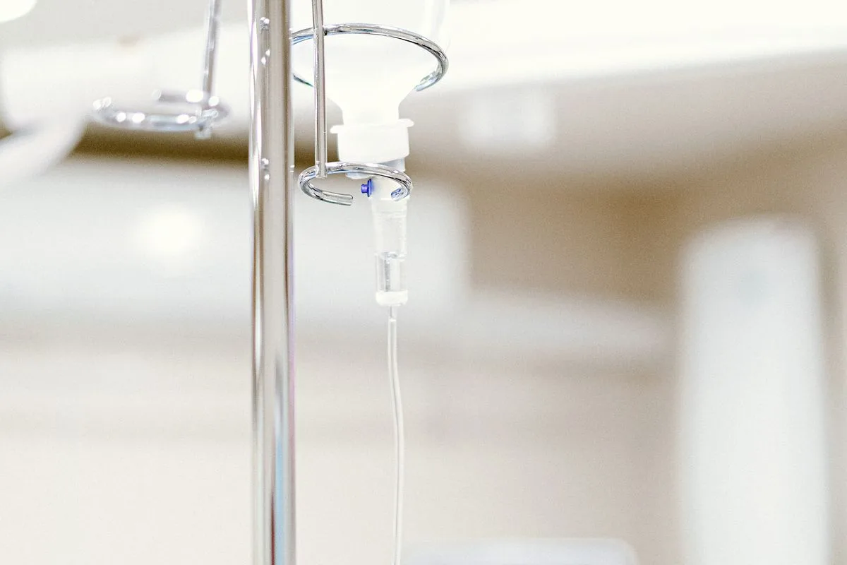
Hysteroscopy is preferred in the investigation of various gynecological complaints and in the treatment of some problems. Since the method offers the opportunity to directly view the inside of the uterus, it takes an important complementary role in evaluating findings that cannot be clearly explained by methods such as ultrasound.
Abnormal or long-term uterine bleeding
Investigation of the cause of infertility
Recurring low story
Intrauterine findings detected on ultrasound
Intrauterine concomitancy suspicion
Each of these conditions may require a different degree of research; in which case the examination and previous examination results are evaluated together to determine if hysteroscopy is appropriate. In some patients, hysteroscopy may not be the first preferred method, but simpler examinations may be requested first.
In postmenopausal abnormal bleeding, hysteroscopy is often a preferred method of evaluation. Detailed examination of the inside of the uterus during this period is important in terms of clarifying the source of the bleeding and sampling if deemed necessary.
In couples with infertility research, hysteroscopy can be used in conjunction with other examinations to assess the structure of the intrauterine cavity and possible barriers. When symptoms such as intrauterine adhesion, septum, or polyp are detected, they can be treated in the same session to prepare the ground for the next steps.
Anesthesia Options in Hysteroscopy
Diagnostic hysteroscopy and minor extensive procedures can often be performed under office conditions with local anesthesia or without any anesthesia at all. This approach facilitates the patient's return to daily life on the same day and eliminates the risks associated with general anesthesia.
In more extensive operative procedures, for example, general or regional anesthesia may be preferred when taking a large polyp or myoma. Which anesthesia method to apply is assessed together in advance based on the scope of the planned procedure and the patient's overall health.
When choosing anesthesia, the patient's anxiety level, pain threshold and responses to previous procedures are also taken into consideration. These preferences are discussed in the pre-consultations with the physician and the most appropriate method is determined together for both the patient's comfort and the safety of the process.
Hysteroscopy Processing Time and Discharge

The processing time varies depending on the scope of the application; While diagnostic hysteroscopy usually lasts a few minutes, operative procedures can take longer. If general anesthesia has not been administered, patients may return to their daily activities after a brief rest following the procedure; Most patients are discharged on the same day.
Vehicle use after discharge is not recommended for that day in patients undergoing general anesthesia; Therefore, it is preferable to return home with the escort. This restriction is usually not the case in procedures performed under office conditions with local anesthesia.
Factors affecting processing time include the size, placement and number of findings identified; Multiple polyps, or a large stickiness, can cause the process to take a little longer. The physician may share a general prediction of this period prior to the procedure.
Post-hysteroscopy Recovery Process
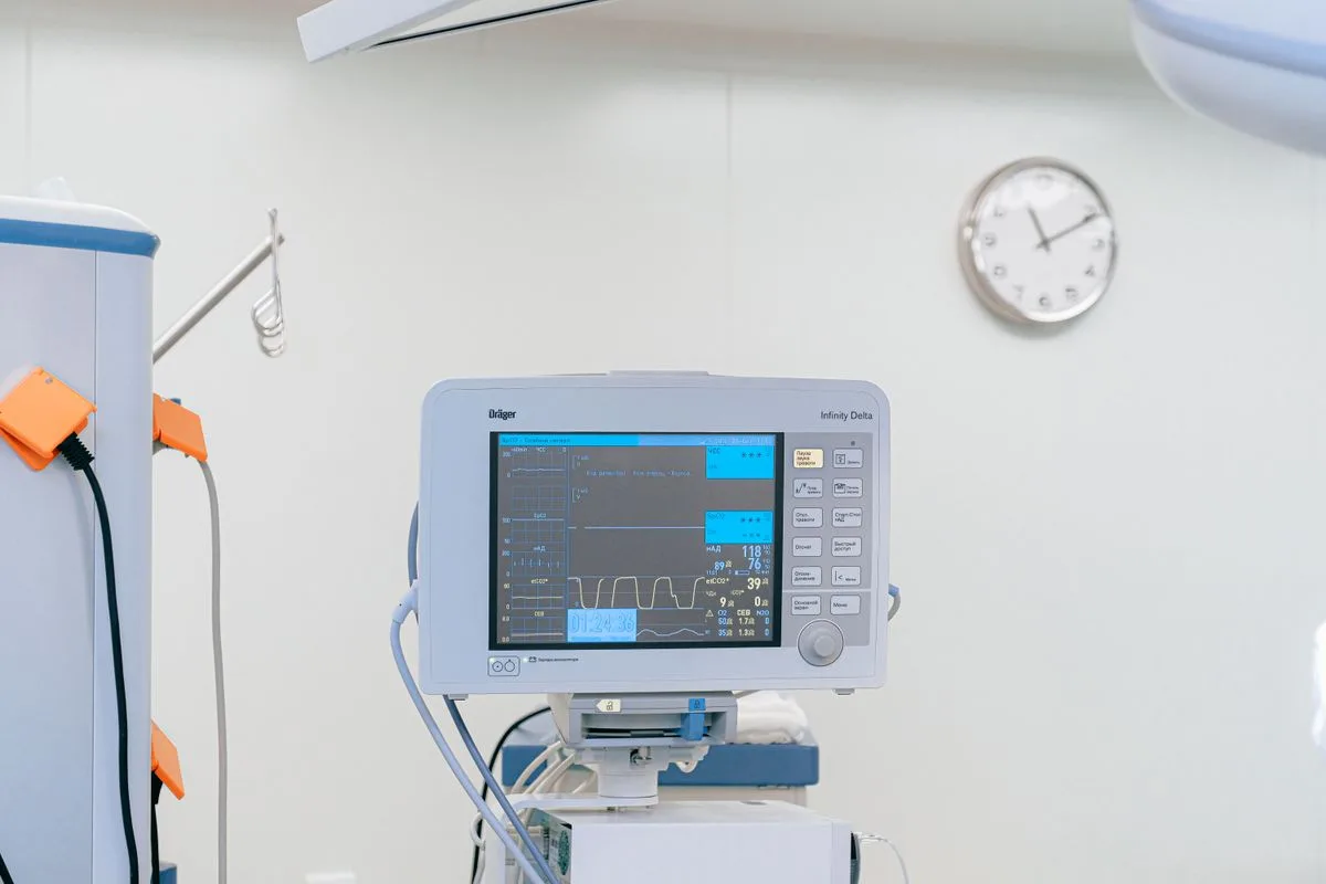
Light cramping and spotting that last for several days after the procedure are common, expected findings. These complaints usually subside spontaneously over time. Simple painkillers recommended by the physician can be used if pain is felt.
A few days of mild cramping and spotting is normal.
It is recommended to avoid severe physical activity for a while.
It is recommended that sexual intercourse be avoided during the period indicated by the physician
If fever, foul-smelling discharge, or severe pain is detected, evaluation should be sought without delay.
The healing process can vary from person to person and depending on the scope of the procedure being implemented. A control examination is valuable in order to confirm that the recovery is progressing as expected and to share an additional recommendation if any.
Taking a shower is usually possible again soon after the procedure; however, tampon use may be asked to avoid during the time recommended by the physician.These recommendations contribute to smooth completion of the recovery process and reduce the risk of infection.
Return to day-to-day work is usually planned gradually; light household chores and desk work may be possible again in most patients within a few days, while the time specified by the physician for tasks requiring heavy lifting may vary depending on the extent of the procedure being performed.
Possible Risks of Hysteroscopy
Hysteroscopy is generally considered a safe method; But as with any medical procedure, it carries some risks. While these risks are rare, prior knowledge helps the patient make informed decisions about the procedure.
Infection
Bleeding
Puncture (perforation) in the uterine wall
Complications due to the expansion fluid used
The frequency of these complications may vary depending on the extent of the procedure and the environment in which it is administered. The physician shares with the patient the possible risks before the procedure and takes a careful approach to minimize these risks during the procedure.
Most of these rare complications are noticed during or immediately after the procedure and can be managed by timely intervention. The physician's experience and the careful approach followed during the procedure play a decisive role in minimizing possible risks.
It is important to contact the physician without wasting time when an unusual finding such as high fever, severe abdominal pain or abundant bleeding is noticed during the post-processing period. Although such findings are rare, early detection helps to plan treatment quickly.
What to Consider Before Hysteroscopy

The medication used and the history of bleeding disorder, if any, should be shared with the physician
Severe physical activity should be avoided the day before the procedure
If general anesthesia is planned, the duration of starvation indicated by the physician must be observed.
It is recommended to plan an escort for a return home after the procedure
These preparatory steps contribute to the safe and smooth progress of the procedure. In the event of any uncertainty, it should not be hesitate to ask the physician during the examination; each patient's preparation process is also evaluated according to his or her own health condition.
It is important to share this condition with the physician if a fever, urinary system infection or vaginal infection is detected within the week before the procedure. In the presence of an active infection, the procedure may be delayed until after recovery.
Patients who take blood thinners may need a temporary adjustment in the dose of the drug in the days before the procedure, directed by the physician. Such changes should always be made under physician's control, the use of medication should not be altered by the patient's own decision.
When Should You Apply to an Expert?
Evaluation should not be delayed when abnormal uterine bleeding, infertility research, recurrent low story or an intrauterine finding detected on ultrasound is noticed. Samsun Gynaecologist The examination by the physician helps to clarify whether hysteroscopy is necessary.
The duration, severity and accompanying findings of complaints play an important role in determining which examinations are a priority. Instead of waiting in a suspicious situation, being examined allows the correct diagnosis and treatment process to begin faster.
The same principles apply to patients who have had previous hysteroscopy and have noticed similar complaints again. Sharing the procedure and its outcome in the past with the physician contributes to the faster evaluation of the current situation.
The Difference Between Hysteroscopy and Laparoscopy
These two methods, often confused by patients, are actually separate endoscopic applications that evaluate different regions. In hysteroscopy, the camera is passed through the mouth of the vagina and uterus to be delivered directly to the intrauterine cavity; No incisions are needed in the abdomen. In laparoscopy, the external surface of the uterus, ovaries, tubes and surrounding pelvic organs are examined by the camera placed in the abdominal cavity through a small hole opening around the belly. In other words, hysteroscopy presents an assessment of the "internal" structure of the uterus, and laparoscopy provides an evaluation of the "outside" appearance of organs inside the abdomen.
This different access path determines the purposes of use of both methods. Hysteroscopy; Intrauterine polyp is preferred in the research and treatment of findings such as myomas that overflow into the concomitant, septum, or uterine cavity, while laparoscopy is mainly used to evaluate endometriosis, ovarian cyst, tube obstruction, or myomas that have settled outside the uterus. Because laparoscopy requires entry into the abdomen, it is often administered under general anesthesia and the healing process may take a little longer compared to hysteroscopy, which is performed without incisions.
In some cases, especially in the study of infertility, both the internal structure and the external environment of the uterus may need to be evaluated together; In such cases, two methods can be planned together in the same session. It is decided at the time of examination which method or two will be appropriate to apply together, taking into account the nature of the complaint and the results of the previous examination.
Conclusion
Samsun hysteroscopy Its method is a reliable approach that makes it possible for the inside of the uterus to be displayed directly and treated in the same session when necessary. Through its diagnostic and operative uses, it finds a wide range of uses in assessing different gynecologic complaints.
Abnormal bleeding, infertility research, or not delaying the examination when a finding detected on ultrasound is important for the correct diagnosis and treatment process to begin on time. Gynecological examination for individual assessment remains the first and most important step in the process.

Original Turkish content prepared and reviewed by
Prof. Dr. Aşkı Ellibeş Kaya
Specialist in Obstetrics and Gynecology
Last updated: 23 August 2026
The content on this page is intended for informational purposes; the diagnosis and treatment decision is made only with your physician after the examination.
References
The following sources relate to the informational issues outlined by them. The personal diagnosis and treatment plan requires physician evaluation. Source texts are in English.
NHS
Hysteroscopy (opens in new tab)Related subject: Camera examination of the uterine cavity, biopsy and polyp removal.
The information on this page is for general informational purposes only and does not replace medical advice, diagnosis or treatment. Each patient's condition is different; the method and process to be applied is determined only by the physician after examination and evaluation.
Frequently asked questions
Diagnostic hysteroscopy and minor extensive procedures are usually completed with a slight feeling of discomfort. Thanks to anesthesia applied in more extensive operative procedures, pain is largely controlled during the procedure. The pain threshold, which varies from person to person, can also affect the experience.
Related Services
Other topics under the topic of gynecology.

Start with an appointment
Let us determine the day and approach that best suits you. Your consultations are conducted on a privacy basis.