

Samsun Hysterosalpingography (HSG)
The hysterosalpingography (HSG) is a diagnostic method that assesses whether the tubes are open with contrasting material and X-ray images with the structure of the uterine cavity. The findings are interpreted together with the physician's examination.
Samsun hysterosalpingography Shooting is an imaging method known as HSG (hysterosalpingography). The structure of the cervix and egg canals are evaluated in this way.
This examination is a method that is frequently used in the diagnosis process of women who have difficulty conception; Imaging is done via X-ray with the help of contrast material. The results lead to further examination and treatment planning; The final assessment is always carried out with a physician's examination. This procedure in Samsun, with expertise in urogynecology and infertility with specialities in women's diseases and childbirth Prof. Dr. Aşkı Ellibeş Kaya It can be evaluated. In the first meeting at the office in Atakum, the patient's story and previous examinations are examined, deciding whether or not hysterosalpingography filming is required accordingly.
What Is Hysterosalpingography (HSG)?
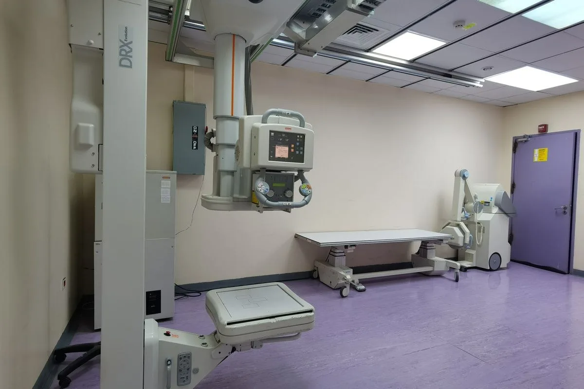
The hysterosalpingography is a radiological diagnostic method called hysterosalpingography (HSG) in the medical language. During the procedure, a special contrast substance is given into the uterus through the cervix; This substance is imaged with an X-ray device to examine whether the egg channels are open by the shape of the uterine cavity. The spread of contrast material into the abdomen is an important finding that the tubes are open.
This examination is usually 5-10 after the end of the menstrual bleeding. It is planned between days. During this period, the images are more clearly obtained as the uterus inner membrane is not yet thickened, and the likelihood of a possible pregnancy is largely excluded. The process is performed by an experienced team at a center that is coordinated with the radiology unit.
The hysterosalpingography is a method that, unlike ultrasonography, can show the internal structure of the cervix and the functional opening of the tubes at the same time. Therefore, it takes a complementary role in infertility assessment along with ultrasonography and hormone tests. Planning the procedure and interpreting the results requires a joint assessment of women's disease and obstetrician and radiology team.
Why Is Hysterosalpingography Performed?
The hysterosalpingography is an important part of infertility research, and is usually brought up in couples who have not been able to obtain pregnancy despite unprotected intercourse for a year (under 35 months). The structural condition of the uterus and tubes must be known before treatments such as IVF or insemination are planned.
Tube obstruction suspicion
Recurring low story
Intrauterine adhesion, polyp or myoma suspicion
Congenital uterine structural anomalies
Routine assessment before IVF or insemination
These findings help the physician determine the treatment roadmap. The results are not evaluated alone, but holistically along with hormone tests, ultrasonography and the patient's history. Sometimes the hysterosalpingography may also be requested to clarify a finding that remains unclear in a previous ultrasonography.
Infertility research, along with the hysterosalpingography, is often done with hormone tests (such as FSH, LH, prolactin, thyroid function tests), ultrasound assessing egg reserve and uterine structure, and a detailed story acquisition and examination. This holistic assessment contributes to the correct determination of the probable cause.
Hysterosalpingography (HSG): Step by Step
The hysterosalpingography process consists of several stages, from pre-date preparation to the outcome report. Knowing how each stage works makes the patient more comfortable during the procedure.
Communicating with the patient at every step of the process is an important element that reduces anxiety level. Especially for the first time, it is recommended that people who will perform this examination share with their physician every detail they are curious about before the procedure.
Appointment and Pre-Prep
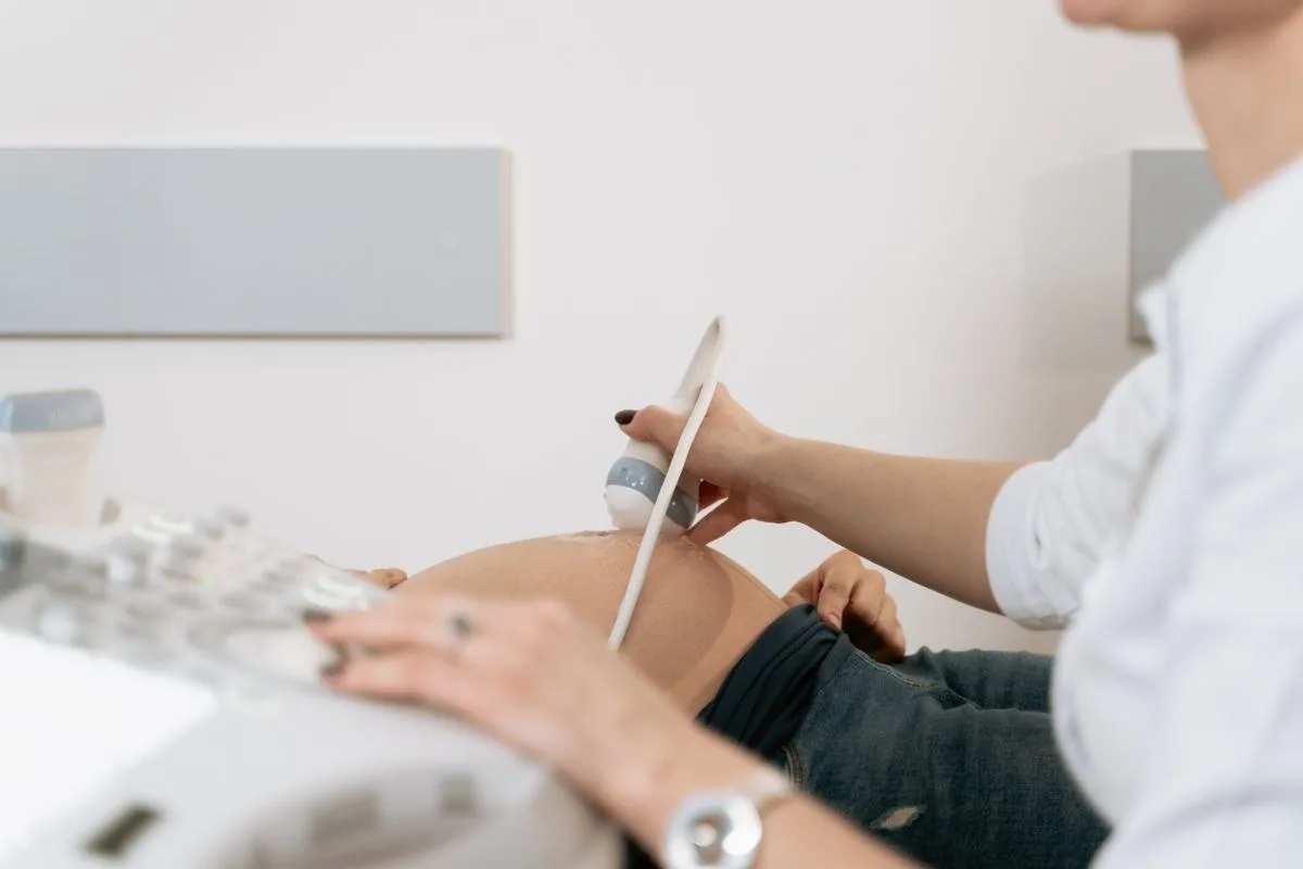
The appointment is usually scheduled according to the appropriate days after the end of the period. If the patient has a urinary tract infection before the procedure, a history of sexually transmitted disease and allergy status are questioned; A simple urine test may be requested if necessary. In some cases, the physician may recommend the use of a short-term painkiller prior to the procedure.
In the first meeting, the physician notes the patient's menstrual pattern, previous pregnancy and low stories, if any, previous surgeries. This information contributes to the planning of the process at the right timing and with the right technique; The patient is encouraged to ask questions and share their concerns at this stage.
The procedure can be performed on a full or hungry stomach; No special dietary restrictions are required. However, it is recommended that the patient come in comfortable clothes and take time for a short rest period after the procedure. It is important to inform the physician in advance about the medications used and chronic diseases.
In the pre-date consultations, information from the patient on the following issues helps to plan the procedure safely:
Chance of conception or date of last menstrual
Known allergy history to contrast substance or iodine
Regularly used drugs and blood thinners
A history of pelvic infection or surgery
Chronic diseases (such as thyroid, diabetes)
What Happens During the Process
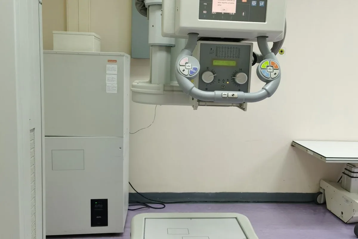
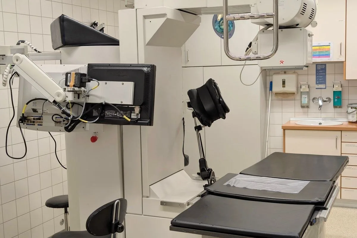
After the patient is placed in the position of a gynecological examination, the cervix is made visible with the help of a speculum. A thin catheter is inserted from the cervix into the uterus and X-ray images are started to be taken with the delivery of contrast material. The transition of contrast material from the tubes to the abdominal cavity is monitored from the screen.
Images are taken from different angles and the shape of the uterine cavity and the course of the tubes are recorded in detail. The team can take additional shots that they deem necessary until the image quality is sufficient, which can extend the processing time by several minutes.
A cramp-like pain may be felt during the procedure; This feeling is usually short-term and varies in severity from person to person. The entire process is completed in an average of 10-15 minutes. Regularly keeping the patient breathing throughout the procedure can help relax the muscles and reduce discomfort.
The hysterosalpingography is not a procedure that requires general anesthesia; The patient's consciousness is clearly made. In patients with a high sensitivity to pain, the physician may recommend reducing the discomfort with a pain reliever taken orally shortly before the procedure. During imaging, the team informs the patient at each stage to ensure that the process proceeds without any surprises.
Result and Report Retrieval
The images are evaluated by the radiologist during the procedure and usually a written report is prepared on the same day or in a short time. This report includes the shape of the uterine cavity, the opening of the tubes, and, if any, noticeable findings.
The report is transmitted to the obstetrician and obstetrician, who is pursuing the patient. Transferring the results to the patient and shaping the treatment plan are carried out in this consultations; Interpretation of technical statements in the report requires physician evaluation.
In this consultations, the patient can direct questions about the report content to his or her physician. If necessary, additional examination or a different imaging method may be planned; this decision is shaped by the nature of the findings and the patient's overall clinical status.
What to Consider When Having Hysterosalpingography
The fact that the hysterosalpingography process is performed in a way that is safe and accurate depends on several basic standards. These standards directly affect both the comfort of the process and the reliability of the results.
Patients learn about the center and team prior to the procedure, allowing them to approach the process more confidently. When making an appointment, it is recommended to clarify issues such as who will perform the process, at which center it will be performed, and when the results will be evaluated.
Expert Doctor and Equipment Standards
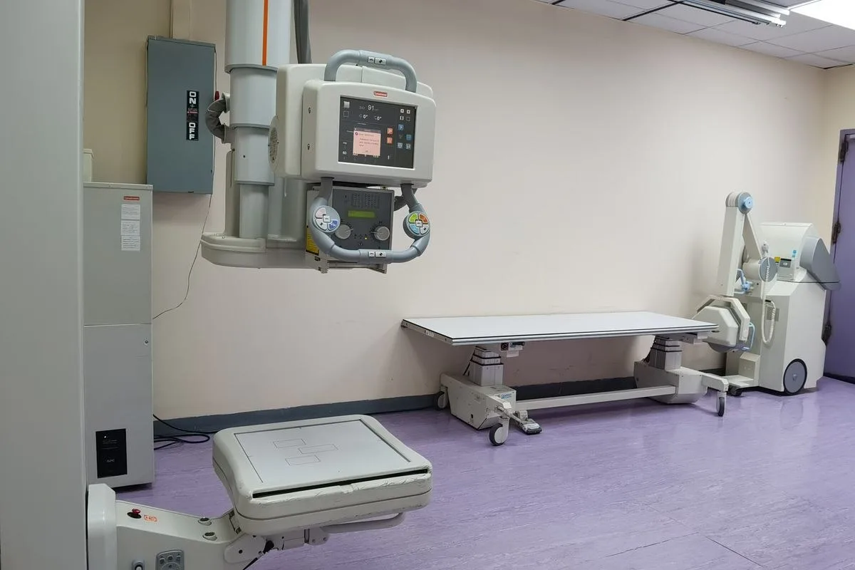
It is important that the procedure be performed in a center with appropriate radiological equipment, under the supervision of a physician with experience in women's diseases and obstetrics. Catheter placement and adjustment of the amount of contrast matter are steps that require experience and can affect image quality when not implemented correctly.
In Samsun, such examinations are carried out in private hospitals and imaging centers contracted by the direction of obstetricians. The physician plans where and when the procedure will be performed according to the patient's history.
The current and regular maintenance of the imaging device ensures that both the image quality and the dose of radiation the patient is exposed to are kept at the lowest possible level. Therefore, it is preferable that the process be performed in a center with the necessary technical infrastructure.
Clinical Hygiene and Sterile Protocol
As with every procedure involving an attempt into the uterus, sterile material use and compliance with hygiene rules are critical to minimizing the risk of infection. Speculum, catheter and other materials used are expected to be disposable or properly sterilized.
If there is an active infection finding (such as discharge, fever, abdominal pain) prior to the procedure, the procedure may be delayed. The physician may recommend preventive medication prior to the procedure if deemed necessary.
Compliance with the sterile protocol is not limited to materials used only; The cleanliness of the processing room, the hands-on hygiene of the staff and the careful technique during catheter placement also directly affect the outcome. The patient may ask his or her physician if they are curious about these matters prior to the procedure.
What to Expect After Hysterosalpingography in Samsun
The mild symptoms after the procedure are mostly temporary. However, some findings need to be carefully followed and consulted with the physician when necessary.
The physician tells the patient verbally expected symptoms and situations that need attention after the procedure is over. This information is important for the patient to be able to follow themselves comfortably at home and apply at the right time when necessary.
Return to Daily Living Activities
Most patients can return to their daily activities within hours of the procedure. On the first day, it is considered normal to experience a feeling of bleeding and cramping in the groin area in the mild staining style. Heavy physical activity and intense sports are recommended to be avoided for a few days after the procedure.
Sexual intercourse is usually recommended to be suspended for 2-3 days; this period may vary according to the physician's assessment. A similar period of stay away from environments such as pool and sea is recommended.
Some of the points recommended to be considered in the post-processing period are:
Restricting the use of painkillers to physician's recommendation
Using a pad instead of a tampon
Consuming plenty of water
Avoiding strenuous movements such as heavy lifting
If recommended by the physician, go to a control examination
Symptoms of Infection and Criteria for Calling a Doctor
In the event of the following symptoms, the physician should be consulted without delay:
fever over 38 degrees
Foul-smelling or coloured vaginal discharge
Increasingly, non-stop abdominal pain
Excessive amounts or prolonged bleeding
General condition disorder such as dizziness, fainting sensation
Although such symptoms are rare, they should not be ignored. Samsun obstetrician Contacting the patient makes it easier for a possible infection to be controlled at an early stage.
If symptoms are mildly observed and tense over time, there may be no mention of an emergency; yet the sharing of every curious situation by telephone or face-to-face with a physician prevents unnecessary anxiety.
How Are Hysterosalpingography Results Interpreted?
The hysterosalpingography report provides detailed information on the shape of the uterine cavity and the state of opening of the tubes. However, it is not enough to interpret the report alone; The physician evaluates the findings along with the patient's clinical chart.
The medical terms used in the report may seem complicated to the patient. Therefore, it is easier for the results to be explained in plain language by the physician and to ask questions until the patient understands.
Normal Hysterosalpingography (HSG) Findings
In a normal hysterosalpingography, the contrasting substance fills the uterine cavity properly and passes freely through both tubes into the abdominal cavity. This appearance indicates that there is no structural barrier to the uterine cavity and that the tubes are open.
The normal result does not mean that the pregnancy is delayed for other reasons; Other elements, such as hormonal factors, egg reserve and male factor, must also be evaluated. Therefore, following the normal hysterosalpingography, the physician continues the couple's overall infertility assessment as a whole.
In couples with normal findings, the physician may make follow-up recommendations to increase the chance of pregnancy by natural means, or consider it appropriate to continue the natural trial for a certain period of time. This decision is personalized based on the couple's age and story.
Abnormal Findings and Treatment Options
Single or double-sided tube obstruction, intrauterine adhesion, polyp, myoma, or congenital uterine deformities are the main abnormal findings that can be detected in the hysterosalpingography. The treatment approach of these findings depends on the type and severity.
The location, size and effect of the finding on the uterus or tubes directly shape the treatment plan. Some findings require only close follow-up, while others may require more detailed examination with additional imaging methods (e.g. ultrasonography or laparoscopy).
Some observations state that mild tube blockages can be opened during the procedure and increase the likelihood of pregnancy within a few months following; However, this effect is not seen in every patient in the same way and it is not right to give a precise rate. In some cases, the problem can be addressed with medication or minor surgical interventions, while in others it can be directed directly to advanced treatment methods such as IVF. Which approach is appropriate is determined by physician examination and additional examinations.
Common Myths and Facts About Hysterosalpingography
There are some misconceptions about the womb film that often circulate among patients. The following table compares scientific facts with the most common myths.
Myth: Fact
The hysterosalpingography permanently damages the ovaries.: There is no evidence that the low-dose X-ray used in the procedure caused permanent damage to the egg reserve.
The procedure is always very painful.: The level of pain varies from person to person; most patients describe a short-term feeling of cramping.
If the result is normal, the pregnancy will be finalized.: The normal result is only related to the structural condition of the uterus and tubes, which does not finalize pregnancy.
Work or school axes for a long time after the procedure.: Most patients may return to their daily routine on the same day or the next.
It can never be withdrawn during menstruation.: During intense bleeding, the procedure is usually delayed, but the timing is determined by the physician.
The most reliable way to get the right information is to meet with the physician who planned the process, which does not replace the personal situation.
Experiences shared on social media or on different patient forums can vary greatly from person to person. Such shares can be read to get an idea; However, the diagnosis and treatment decision should only be based on examination and examination results.
Conclusion
Samsun hysterosalpingography The shot is a relatively short-lived study, which provides important information for women who are planning pregnancy and are in the process of diagnosis. Proper timing, an experienced team, and sterile conditions play a decisive role in the safe completion of the process.
Every subject that is curious before and after the procedure should be evaluated with the physician in person. Contacting the practice in Samsun Atakum for appointment and pre-consultations ensures that the process is planned in the most accurate way.
The findings after the womb film are only part of the couple's overall infertility assessment. A permanent treatment decision is shaped holistically by the physician, along with hormone tests and examination findings.

Original Turkish content prepared and reviewed by
Prof. Dr. Aşkı Ellibeş Kaya
Specialist in Obstetrics and Gynecology
Last updated: 28 August 2026
The content on this page is intended for informational purposes; the diagnosis and treatment decision is made only with your physician after the examination.
References
The following sources relate to the informational issues outlined by them. The personal diagnosis and treatment plan requires physician evaluation. Source texts are in English.
NHS
Diagnosis of infertility (opens in new tab)Related subject: Imaging of uterine and tube openings with HSG.
The information on this page is for general informational purposes only and does not replace medical advice, diagnosis or treatment. Each patient's condition is different; the method and process to be applied is determined only by the physician after examination and evaluation.
Frequently asked questions
Most patients experience a short-term cramping sensation, with the level of pain varying from person to person. The discomfort usually does not last long, as the process is completed within a few minutes.
Related Services
Other issues under the title IVF.



Start with an appointment
Let us determine the day and approach that best suits you. Your consultations are conducted on a privacy basis.